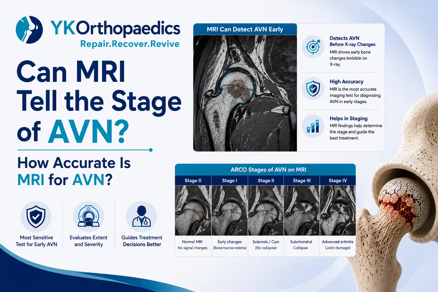
MRI can provide the information needed to stage AVN very accurately, particularly in the early stages, but the MRI report alone should not always be treated as the final stage.
Doctors usually look at:
- Whether AVN is present
- Size of the lesion
- Location of the lesion
- Weight-bearing surface involvement
- Subchondral fracture
- Femoral-head collapse
- Cartilage and joint damage
MRI is highly sensitive for early AVN, but determining the exact stage requires interpretation of the complete imaging findings using an appropriate staging system such as Ficat or ARCO.
How Accurate Is MRI for Detecting AVN?
MRI is considered one of the most sensitive imaging methods for detecting osteonecrosis, especially during the early stages.
It can identify bone changes that may not yet appear on an X-ray. This makes MRI particularly useful when:
- Hip pain is unexplained.
- AVN is suspected because of risk factors.
- X-rays are normal but symptoms continue.
- AVN is already present in the opposite hip.
- The doctor needs to assess the extent of disease.
In other words:
Normal X-ray does not always rule out early AVN.
MRI can provide much earlier information about the condition of the bone.
Can MRI Detect AVN Before Symptoms Become Severe?
Yes.
Early AVN can produce few symptoms or even no symptoms, while MRI may already show characteristic changes within the bone.
This is important because treatment aimed at preserving the natural joint is generally more feasible before significant femoral-head collapse develops.
Therefore, the severity of pain does not necessarily tell you the stage of AVN.
For example:
A patient may have:
Mild pain + significant AVN on MRI
or
Severe pain + relatively early AVN
This is why imaging and clinical examination are both important.
Can MRI Tell the Exact Stage of AVN?
MRI can help determine the stage, but the exact stage depends on the staging system used.
Several systems are used to classify femoral-head AVN, including:
- Ficat classification
- ARCO classification
- Other imaging-based classification systems
These systems do not always define stages in exactly the same way.
Therefore, an MRI report may describe the findings without explicitly saying:
"Stage 2 AVN."
Your orthopedic specialist interprets the findings and applies the relevant staging system.
How Does MRI Help Identify Different AVN Stages?
A simple way to understand the progression is:
| AVN Stage | Typical Imaging Findings |
|---|---|
| Stage 1 | MRI abnormalities may be present while X-ray can still be normal |
| Stage 2 | AVN-related bone changes are visible, but significant collapse is absent |
| Stage 3 | Subchondral fracture and/or femoral-head collapse develops |
| Stage 4 | Collapse is accompanied by secondary degenerative joint changes/arthritis |
This is a simplified explanation because the exact definitions vary between classification systems.
What Does MRI Show in Stage 1 AVN?
Stage 1 is an early phase.
MRI may show:
- Abnormal bone marrow signal
- Characteristic AVN pattern
- Serpiginous/geographic lesion
- Possible double-line sign
- No significant femoral-head collapse
X-rays may still appear normal at this stage.
Why is this important?
Early detection may provide a better opportunity to consider joint-preserving treatment in appropriately selected patients.
What Does MRI Show in Stage 2 AVN?
Stage 2 generally means that AVN-related structural changes are visible, but significant collapse has not yet occurred.
MRI may show:
- Clearly defined AVN lesion
- Abnormal bone marrow signal
- Serpiginous margins
- Double-line sign
- No major femoral-head collapse
At this stage, the size and location of the lesion become particularly important.
A small lesion outside the main weight-bearing region may have a different prognosis from a large lesion involving the weight-bearing surface.
What Does MRI Show in Stage 3 AVN?
Stage 3 represents structural failure.
MRI may show:
- Subchondral fracture
- Crescent sign
- Early femoral-head collapse
- Change in femoral-head contour
The presence of a subchondral fracture or collapse is particularly important because it means the weakened bone has started to fail structurally.
What Does MRI Show in Stage 4 AVN?
Advanced AVN can show:
- Significant femoral-head collapse
- Flattening or deformity
- Cartilage damage
- Joint-space narrowing
- Secondary osteoarthritis
- Other degenerative joint changes
At this stage, treatment often shifts from trying to preserve the damaged bone toward restoring the function of the damaged joint.
For patients with substantial collapse and joint degeneration, total hip replacement may be considered.
Which MRI Finding Is Most Important for Staging AVN?
There is no single MRI finding that determines every patient's stage.
However, one of the most important questions is:
Has the femoral head collapsed?
The difference between:
AVN without collapse
and
AVN with subchondral fracture/collapse
can significantly change the treatment approach.
Doctors also consider:
- Lesion size
- Lesion location
- Weight-bearing involvement
- Cartilage condition
- Joint-space changes
- Symptoms
What Does "No Collapse" Mean on MRI?
If your MRI says:
"No evidence of femoral-head collapse."
it generally means that significant structural collapse has not been identified.
This can be an important finding because joint-preserving treatment is generally more feasible before substantial collapse develops.
However, it does not mean that the AVN is harmless.
A large lesion involving the weight-bearing surface may still have a considerable risk of progression.
What Does "Subchondral Fracture" Mean?
A subchondral fracture is a fracture occurring beneath the cartilage-covered surface of the joint.
In AVN, it indicates that the weakened bone has begun to fail.
If the report mentions:
- "Subchondral fracture"
- "Crescent sign"
- "Subchondral collapse"
the finding should be discussed with an orthopedic specialist because it suggests more advanced structural disease.
What Does the Crescent Sign Mean?
The crescent sign is an imaging indication of subchondral fracture beneath the joint surface.
It is important because it indicates that the weakened femoral head has started to undergo structural failure.
It can therefore be an important factor when distinguishing earlier AVN from disease that has progressed toward collapse.
Can MRI Show How Large the AVN Lesion Is?
Yes.
MRI can show the extent of the affected bone and help doctors assess the size and location of the lesion.
This is important because lesion size can influence prognosis.
Small lesion
A small lesion may have a lower risk of collapse, particularly when it does not involve the main weight-bearing region.
Large lesion
A large lesion, particularly one involving the weight-bearing surface, can have a higher risk of structural collapse.
Can MRI Show Whether the Weight-Bearing Area Is Involved?
Yes.
MRI can help identify whether AVN involves the portion of the femoral head that carries significant load during standing and walking.
This is an important factor because lesions in weight-bearing regions experience repeated mechanical stress.
Therefore:
Small + non-weight-bearing lesion
may have a different prognosis from:
Large + weight-bearing lesion.
Can MRI Predict Whether AVN Will Collapse?
MRI can help estimate the risk, but it cannot guarantee exactly what will happen.
The risk of progression depends on several factors, including:
- Size of the lesion
- Location
- Weight-bearing involvement
- Stage
- Underlying cause
- Patient-specific factors
Larger lesions involving weight-bearing areas generally have a higher risk of collapse than small lesions outside those areas.
Is MRI More Accurate Than X-Ray for AVN?
For early AVN, MRI is generally much more sensitive than X-ray.
X-Ray
Useful for:
- Detecting established structural changes
- Assessing collapse
- Monitoring advanced disease
But early AVN may not be visible.
MRI
Useful for:
- Detecting early AVN
- Identifying the lesion
- Assessing its extent
- Detecting subchondral fracture
- Evaluating collapse
- Assessing the opposite hip
Therefore, a normal X-ray does not necessarily exclude early AVN when clinical suspicion remains high.
Does Every MRI Showing AVN Mean I Need Treatment?
No.
The treatment decision depends on:
- Stage
- Lesion size
- Lesion location
- Symptoms
- Risk factors
- Presence or absence of collapse
- Condition of the joint
Small, early lesions may sometimes be monitored or managed conservatively, while selected patients may be considered for joint-preserving procedures.
Once significant collapse and arthritis develop, joint replacement may become more appropriate.
Can MRI Help Decide Whether Core Decompression Is Suitable?
Yes, MRI findings are an important part of the decision.
Core decompression is generally considered for selected patients with early-stage AVN before significant femoral-head collapse.
MRI can help the surgeon determine:
- Size of the lesion
- Location
- Stage
- Whether collapse is present
- Whether the weight-bearing area is involved
This information helps determine whether joint-preserving treatment is appropriate.
Can MRI Tell If I Need a Hip Replacement?
MRI can provide important information for this decision, but MRI alone does not determine whether you need hip replacement.
The orthopedic surgeon also considers:
- Severity of pain
- Walking ability
- Range of motion
- Degree of collapse
- Arthritis
- Age
- Activity level
- Overall health
If substantial femoral-head collapse and secondary arthritis are present, total hip replacement may be considered.
Can MRI Show AVN in Both Hips?
Yes.
MRI can identify AVN in both femoral heads.
This can be particularly useful because AVN may be bilateral even when symptoms are mainly present on one side.
For example:
Right hip: Stage 3 AVN
Left hip: Early AVN without collapse
The treatment plan can therefore be different for each hip.
What If My MRI Says "Bilateral AVN"?
Bilateral AVN means that both hips show evidence of osteonecrosis.
It does not necessarily mean that both hips have the same severity.
You should look at the report for:
- Size of the lesion
- Stage
- Collapse
- Subchondral fracture
- Weight-bearing involvement
- Joint degeneration
Each hip should be evaluated individually.
Can MRI Be Wrong About AVN?
MRI is highly sensitive for detecting AVN, but no imaging test should be interpreted in isolation.
Certain MRI findings can overlap with other bone conditions, and the final diagnosis should take into account:
- Symptoms
- Medical history
- Risk factors
- Physical examination
- MRI findings
- X-ray or other imaging when appropriate
The radiologist's interpretation and orthopedic assessment are therefore important.
Why Can the MRI Report and Doctor's Stage Be Different?
This can happen because the radiologist may describe imaging findings while the orthopedic surgeon applies a particular staging system to the complete clinical picture.
For example, a report might say:
"AVN of the femoral head with no evidence of collapse."
The orthopedic surgeon may then classify it according to a specific system and determine the treatment based on lesion size and location.
So, if your MRI report does not explicitly mention "Stage 2" or "Stage 3," that does not mean the report is incomplete.
What Should You Look for in Your AVN MRI Report?
If you have the report in front of you, look for these terms:
Signs suggesting earlier disease
- AVN/osteonecrosis
- Serpiginous lesion
- Geographic lesion
- Double-line sign
- No subchondral fracture
- No femoral-head collapse
Signs suggesting progression
- Subchondral fracture
- Crescent sign
- Femoral-head collapse
- Flattening
- Deformity
Signs suggesting advanced joint damage
- Cartilage loss
- Joint-space narrowing
- Secondary osteoarthritis
- Degenerative changes
These findings can help you understand the report, but the final stage should be confirmed by your orthopedic specialist.
What Happens After an MRI Diagnosis of AVN?
Treatment depends on the stage and structural condition of the hip.
Early AVN Without Collapse
Possible approaches include:
- Activity modification
- Protected weight-bearing
- Pain management
- Physiotherapy
- Monitoring
- Joint-preserving treatment in selected patients
AVN With Significant Collapse
Once substantial collapse and joint degeneration develop, joint-preserving treatment becomes less predictable.
Total hip replacement may be considered when pain and functional limitations are significant.
The goal is to relieve pain and restore mobility.
When Should You See an Orthopedic Doctor?
You should seek orthopedic evaluation if your MRI shows AVN and you have:
- Persistent hip or groin pain
- Increasing pain while walking
- Difficulty climbing stairs
- Hip stiffness
- Reduced range of motion
- Limping
- Subchondral fracture
- Femoral-head collapse
- Bilateral AVN
- A history of long-term steroid use
- Previous hip trauma
Early assessment can be particularly valuable because treatment options aimed at preserving the natural joint are generally more useful before significant collapse.
Expert Opinion
According to Dr. Yugal Karkhur, a Best Orthopedic Doctor in Gurgaon, MRI is one of the most useful investigations for understanding AVN because it can detect the disease early and show the extent of bone involvement.
However, the MRI should be interpreted by looking at the complete picture—not just the word "AVN." Lesion size, location, weight-bearing involvement, subchondral fracture and femoral-head collapse are particularly important when assessing the stage and treatment options.
Early AVN without collapse may offer an opportunity for joint-preserving treatment in selected patients. When significant collapse and secondary arthritis have developed, treatment may shift toward hip replacement.
Conclusion
MRI is one of the most accurate and useful imaging tests for detecting AVN, especially during its early stages. It can show the location and extent of the lesion and identify important changes such as subchondral fracture and femoral-head collapse.
MRI can provide the information needed to stage AVN, but the exact stage depends on the classification system being used and the combination of imaging findings. A report should therefore be interpreted by an orthopedic specialist rather than by looking at one phrase in isolation.
The most important questions are:
Is AVN present? → How large is it? → Is the weight-bearing area involved? → Is there a subchondral fracture? → Has the femoral head collapsed? → Is there joint damage?
If you are looking for a Best Orthopedic Doctor in Gurgaon, Dr. Yugal Karkhur can evaluate your MRI findings, determine the extent and stage of AVN and discuss appropriate treatment options.
FAQs
Can MRI detect AVN early?
Yes. MRI is highly sensitive for early osteonecrosis and can detect changes before they become visible on an X-ray.
Can MRI tell what stage of AVN I have?
MRI can provide the imaging information needed for staging, but the exact stage depends on the classification system used and interpretation of the complete findings.
Is MRI better than X-ray for early AVN?
Yes. MRI is generally much more sensitive for detecting early AVN, while X-rays are useful for identifying established structural changes and collapse.
What MRI finding indicates advanced AVN?
Subchondral fracture, crescent sign, femoral-head collapse and secondary degenerative changes indicate progressively advanced disease.
Does "no collapse" mean my AVN is early?
It generally suggests that significant structural collapse has not occurred, but the exact stage also depends on the other imaging findings and the staging system being used.
Can MRI show the size of an AVN lesion?
Yes. MRI can help determine the extent and location of the affected bone, which can be important for assessing prognosis and treatment options.
Can MRI show whether AVN affects the weight-bearing area?
Yes. MRI can help identify whether the lesion involves the weight-bearing portion of the femoral head, an important factor when assessing the risk of collapse.
Does every MRI-diagnosed AVN require surgery?
No. Treatment depends on the stage, lesion characteristics, symptoms and joint condition. Selected early cases may be managed conservatively or with joint-preserving treatment.
Can MRI tell whether I need hip replacement?
MRI provides important information, but the decision also depends on pain, mobility, examination, degree of collapse, arthritis and overall health.
Can MRI detect AVN in both hips?
Yes. MRI can identify AVN in both femoral heads, even when symptoms are more noticeable on only one side.