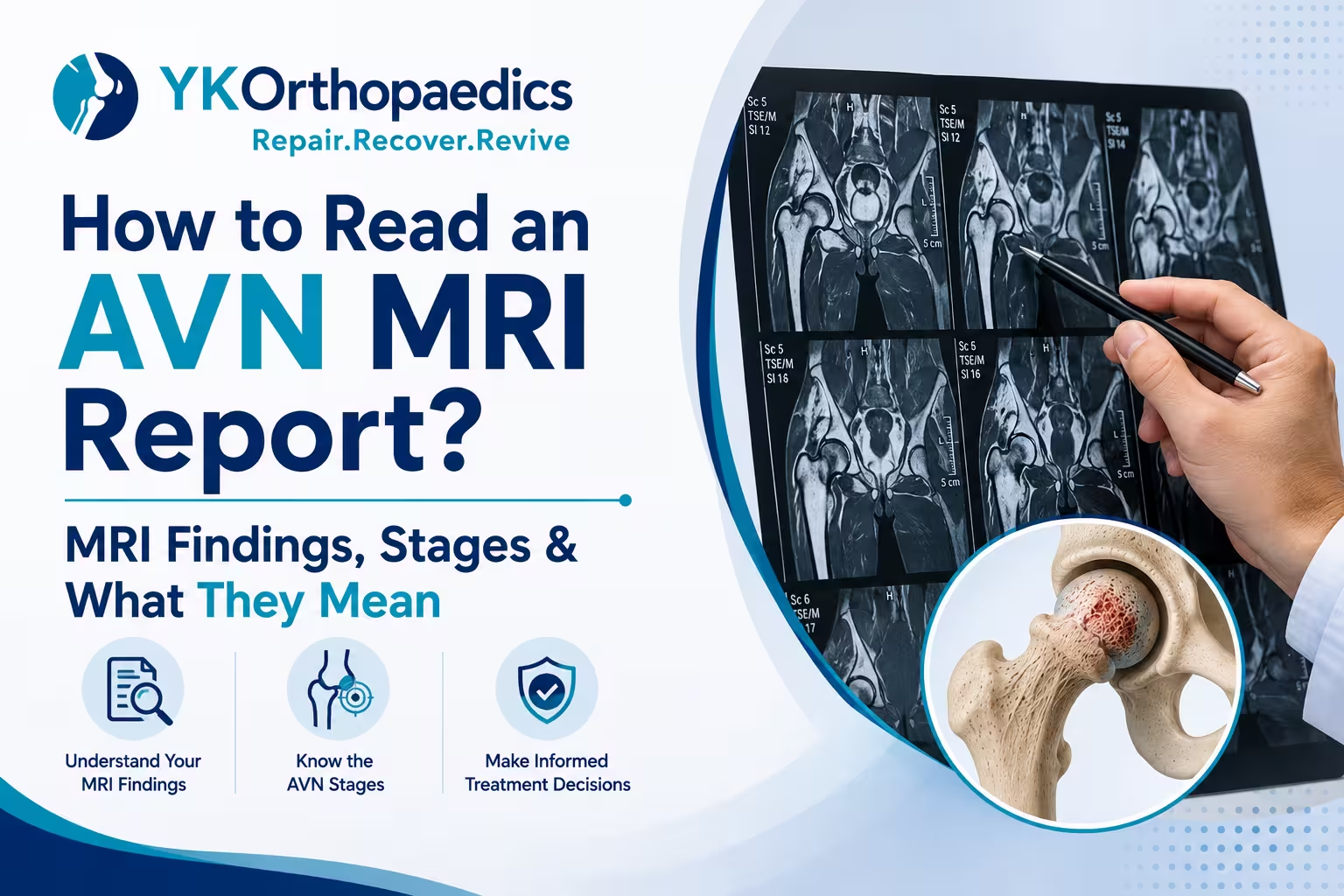
An AVN (Avascular Necrosis) MRI report helps determine whether osteonecrosis is present, where it is located, how much bone is affected and whether the bone has started to collapse. MRI is particularly important because it can detect AVN earlier than an X-ray, sometimes before significant symptoms or visible X-ray changes develop.
When reading an AVN MRI report, some of the most important terms to understand are bone marrow signal changes, serpiginous margins, double-line sign, subchondral fracture, femoral-head collapse, lesion size and joint-space or cartilage changes. However, an MRI finding should not be interpreted in isolation. The actual AVN stage depends on the imaging pattern, extent of involvement and whether collapse or arthritis is present.
What Does an AVN MRI Report Usually Tell You?
An MRI report may provide information about:
- Presence or absence of AVN
- Which hip or bone is affected
- Size and location of the lesion
- Whether the weight-bearing area is involved
- Bone marrow changes
- Subchondral fracture
- Femoral-head collapse
- Joint-space changes
- Cartilage damage
- Bone marrow edema
- Fluid in the joint
- Whether the opposite hip is affected
These details help the orthopedic specialist determine the stage and treatment approach.
Common AVN MRI Terms and Their Meaning
1. "Avascular Necrosis" or "Osteonecrosis"
If your report says:
"Findings suggestive of AVN"
it means the MRI shows changes consistent with loss of blood supply and bone tissue damage.
AVN is also called osteonecrosis, aseptic necrosis or ischemic necrosis of bone.
2. "Femoral Head AVN"
This means the AVN is affecting the ball-shaped upper end of the thigh bone, which forms the hip joint.
The femoral head is the most commonly affected location of osteonecrosis.
For example:
"AVN of bilateral femoral heads"
means AVN is present in both hip joints.
3. "Bone Marrow Signal Changes"
MRI detects changes in the bone marrow that may not yet be visible on X-ray.
Your report may mention:
- Abnormal marrow signal
- Altered marrow intensity
- Bone marrow edema
- Geographic marrow abnormality
These findings need to be interpreted together with the other MRI features rather than being considered diagnostic of a particular stage by themselves.
MRI can detect early osteonecrosis because it is highly sensitive to changes within the bone marrow.
4. "Serpiginous" or "Geographic" Lesion
You may see wording such as:
"Serpiginous lesion involving the femoral head."
A serpiginous pattern describes a winding or irregular border around the affected bone.
This type of imaging description can be seen with osteonecrosis and helps the radiologist identify the affected area.
5. "Double-Line Sign"
The double-line sign is a characteristic MRI finding associated with osteonecrosis.
It refers to two distinct signal lines at the margin of the affected bone on certain MRI sequences.
If your report says:
"Double-line sign noted"
it supports the diagnosis of AVN, but the phrase alone does not tell you whether you have Stage 1, Stage 2, Stage 3 or Stage 4 disease.
The stage also depends on whether structural collapse or arthritis is present.
6. "Subchondral Fracture"
This is one of the most important terms in an AVN MRI report.
A subchondral fracture means there is a fracture just beneath the joint surface.
If your report says:
"Subchondral fracture is noted."
or
"Crescent sign is present."
it indicates structural failure of the affected bone.
This is associated with more advanced AVN and is an important distinction from earlier disease without collapse.
7. "Crescent Sign"
The crescent sign refers to a subchondral fracture that can develop in the femoral head as AVN progresses.
It is an important imaging indicator because it suggests that the weakened bone beneath the joint surface has started to fail.
If your report mentions a crescent sign, you should discuss the finding with an orthopedic specialist.
8. "Femoral Head Collapse"
This is another major term to look for.
If the report says:
"No evidence of femoral-head collapse."
that generally means the joint surface has not undergone significant structural collapse.
If it says:
"Femoral-head collapse is noted."
the disease has progressed to a more advanced structural stage.
Collapse can change the shape of the femoral head and eventually contribute to cartilage damage and secondary osteoarthritis.
9. "Flattening of Femoral Head"
If your report says:
"Flattening of the femoral head"
it means the normally rounded femoral head has lost some of its shape.
This can occur after structural collapse and is generally associated with more advanced AVN.
10. "Secondary Osteoarthritis"
You may see terms such as:
- Secondary osteoarthritis
- Degenerative changes
- Joint-space narrowing
- Cartilage loss
- Osteophytes
- Articular surface irregularity
These findings suggest that AVN has progressed beyond the bone itself and has begun affecting the joint.
Advanced osteonecrosis can lead to secondary osteoarthritis after collapse of the affected bone.
11. "Joint Space Maintained"
If your report says:
"Joint space is maintained."
this generally means there is no significant narrowing of the joint space on the imaging being described.
It is useful information, but joint-space preservation alone does not determine the AVN stage.
The radiologist and orthopedic surgeon also consider the lesion, subchondral fracture and degree of collapse.
12. "No Articular Surface Collapse"
This is generally a favorable finding.
It means that although AVN may be present, there is no significant collapse of the joint surface identified on the MRI.
This distinction is important because treatment options aimed at preserving the natural joint are generally more useful before major collapse occurs.
13. "Bone Marrow Edema"
Bone marrow edema means there is increased fluid-related signal within the bone marrow.
It can occur with several different bone and joint conditions, so bone marrow edema by itself does not prove that AVN is advanced.
The radiologist interprets it together with the characteristic AVN pattern and other findings.
How MRI Findings Relate to AVN Stages
The exact staging terminology can vary because different systems, including Ficat and ARCO, are used to classify femoral-head osteonecrosis.
For a simple patient-friendly understanding:
| AVN Stage | Typical MRI/Imaging Meaning |
|---|---|
| Stage 1 | Early AVN; X-ray may be normal; MRI detects abnormal bone changes |
| Stage 2 | Structural bone changes are visible but no significant collapse |
| Stage 3 | Subchondral fracture and/or femoral-head collapse |
| Stage 4 | Collapse with advanced joint degeneration/secondary arthritis |
This is a simplified explanation, not a substitute for the specific staging system used in your report.
Stage 1 AVN MRI Findings
In early AVN:
- X-ray may be normal.
- MRI may show characteristic AVN changes.
- There is no significant femoral-head collapse.
- Joint structure may still be relatively preserved.
This is why MRI is particularly valuable when AVN is suspected but the X-ray is normal.
What does it mean?
Stage 1 generally represents an early opportunity to consider joint-preserving treatment, depending on lesion size and location.
Stage 2 AVN MRI Findings
Stage 2 generally means:
- AVN is clearly visible.
- Bone structural changes are present.
- The femoral head remains relatively intact.
- There is no major collapse.
The size and location of the lesion are important when estimating the risk of progression.
What does it mean?
Selected patients may be considered for joint-preserving treatment before collapse develops.
Stage 3 AVN MRI Findings
Stage 3 is characterized by structural failure.
The report may mention:
- Subchondral fracture
- Crescent sign
- Early collapse
- Femoral-head contour abnormality
What does it mean?
This is a more advanced stage because the bone has started to fail structurally.
The treatment approach depends heavily on how much collapse has occurred.
Stage 4 AVN MRI Findings
Advanced AVN may show:
- Significant femoral-head collapse
- Flattening or deformity
- Cartilage damage
- Joint-space narrowing
- Secondary osteoarthritis
- Degenerative changes
What does it mean?
When advanced collapse and arthritis are present, joint replacement may become the most reliable treatment option, particularly when pain and loss of function are significant.
What Does "No Collapse" Mean in an AVN MRI?
If your MRI says:
"No evidence of femoral-head collapse."
this generally means the bone has not undergone significant structural collapse.
This is an important finding because treatment aimed at preserving the natural joint is generally more feasible before collapse occurs.
However, "no collapse" does not mean the AVN is harmless or guaranteed to remain stable.
The lesion size and location still matter.
What Does "Extensive AVN" Mean?
If the report describes:
"Extensive AVN"
it means a relatively large area of bone is affected.
The exact significance depends on:
- Percentage of the femoral head involved
- Location of the lesion
- Whether it involves the weight-bearing region
- Presence or absence of collapse
A larger lesion involving a major weight-bearing region generally has a greater risk of progression.
What Does "Weight-Bearing Surface Involvement" Mean?
This is an important phrase in an AVN report.
The weight-bearing portion of the femoral head experiences substantial forces during standing and walking.
If AVN involves this area, the risk of structural collapse may be greater than with a small lesion outside the main weight-bearing region.
Therefore, don't look only at the word "AVN" in the report—look at where the lesion is located.
What Does "Bilateral AVN" Mean on MRI?
If your report says:
"Bilateral femoral-head AVN"
it means both hips are affected.
Importantly, the severity can be different on each side.
For example:
Right hip: AVN without collapse
Left hip: AVN with subchondral fracture and collapse
The treatment plan may therefore be different for each hip.
In non-traumatic AVN, doctors may evaluate both hips because disease can be bilateral.
Does MRI Tell the Exact AVN Stage?
MRI provides critical information for staging, but the final stage should be determined by the orthopedic specialist using the complete imaging findings and the staging system being applied.
A report may describe findings without explicitly writing:
"Stage 2 AVN."
For example, your report might say:
"Serpiginous signal abnormality in the femoral head with no evidence of subchondral collapse."
The doctor uses these findings to determine the stage and treatment implications.
Can I Determine My AVN Stage From the MRI Report Alone?
You can get a general idea, but you should not self-diagnose the stage solely from individual phrases.
For example:
"AVN with no collapse"
→ suggests earlier disease.
"Subchondral fracture/crescent sign"
→ suggests structural progression.
"Femoral-head collapse with secondary osteoarthritis"
→ indicates advanced disease.
But the complete MRI, lesion size, location and clinical examination matter.
What MRI Findings Are Most Important for Treatment?
If you have an AVN MRI report, pay particular attention to:
1. Presence of AVN
Is osteonecrosis actually present?
2. Lesion Size
How much of the femoral head is affected?
3. Lesion Location
Is the weight-bearing portion involved?
4. Subchondral Fracture
Is there a fracture beneath the joint surface?
5. Collapse
Has the femoral head started to flatten or collapse?
6. Arthritis
Is there cartilage damage or secondary osteoarthritis?
These factors help determine whether the focus should be on joint preservation or joint replacement.
Can an MRI Predict Whether AVN Will Get Worse?
MRI provides important information about the extent and structural status of AVN, but it cannot guarantee exactly how an individual patient's disease will progress.
Small lesions may remain stable or heal, while larger lesions—particularly those involving weight-bearing areas—have a greater risk of collapse.
Regular follow-up may therefore be recommended depending on the individual case.
What Treatment Does an AVN MRI Result Lead To?
Treatment depends on the stage, lesion characteristics, symptoms and overall condition of the joint.
Early AVN Without Collapse
Possible approaches include:
- Activity modification
- Protected weight-bearing
- Pain management
- Physiotherapy
- Joint-preserving procedures in selected patients
AVN With Significant Collapse
When the bone has substantially collapsed and the joint has developed advanced degeneration, joint replacement may be required.
Core decompression is generally used for selected earlier-stage disease rather than advanced collapsed joints.
Can You Send Your AVN MRI Report to a Doctor?
Yes, and it can be very useful.
If you have an MRI report, your orthopedic specialist will usually consider both:
The written report + actual MRI images + symptoms + examination
rather than relying on the written report alone.
If you are reviewing your report yourself, do not panic when you see words such as "necrosis," "marrow edema" or "lesion." These terms need to be interpreted in context.
Expert Opinion
According to Dr. Yugal Karkhur, a Orthopedic Doctor in Gurgaon, an AVN MRI report should be interpreted by looking beyond the simple presence of "AVN." The most important questions are how large the lesion is, where it is located, whether the weight-bearing area is involved and, most importantly, whether there is subchondral fracture or femoral-head collapse.
Early diagnosis before significant collapse can provide more opportunities for joint-preserving treatment. Once substantial collapse and joint degeneration have developed, treatment may shift toward joint replacement.
Conclusion
Reading an AVN MRI report becomes easier when you focus on a few key findings: location and size of the AVN lesion, weight-bearing involvement, subchondral fracture, femoral-head collapse and secondary arthritis.
MRI is particularly valuable because it can identify AVN earlier than X-rays and help doctors assess the extent of bone damage.
In simple terms, AVN without collapse generally represents earlier disease, while a subchondral fracture or femoral-head collapse indicates more advanced structural damage. Once significant collapse and arthritis develop, joint replacement may become necessary.
If you are looking for a Best Orthopedic Doctor in Gurgaon, Dr. Yugal Karkhur can review your MRI findings, assess the stage and extent of AVN and discuss the appropriate treatment options.
FAQs
What does AVN mean on an MRI?
AVN means that the MRI shows changes consistent with avascular necrosis or osteonecrosis—damage to bone associated with disrupted blood supply.
What does "no collapse" mean in an AVN MRI?
It generally means that significant structural collapse of the affected bone has not been identified. This is an important distinction because joint-preserving treatment is generally more feasible before major collapse.
What does the double-line sign mean in AVN?
The double-line sign is a characteristic MRI finding associated with osteonecrosis. It supports the diagnosis but does not, by itself, determine the exact AVN stage.
What does a crescent sign mean?
A crescent sign indicates a subchondral fracture beneath the joint surface and suggests structural failure of the affected bone.
Is bone marrow edema the same as AVN?
No. Bone marrow edema can occur in several conditions. It needs to be interpreted together with the other MRI findings.
Can an MRI show Stage 1 AVN?
Yes. MRI is highly sensitive for detecting early osteonecrosis and can identify AVN when an X-ray is still normal.
Does AVN on MRI always mean I need hip replacement?
No. Treatment depends on the stage, lesion size and location, presence of collapse, symptoms and condition of the joint. Early disease may have joint-preserving treatment options.
What is the most important finding in an AVN MRI report?
One of the most important questions is whether subchondral fracture or femoral-head collapse has occurred. Lesion size and location are also important for assessing prognosis and treatment options.
Can both hips show AVN on MRI?
Yes. AVN can affect both hips, particularly in non-traumatic cases. The stage and severity can be different in each hip.