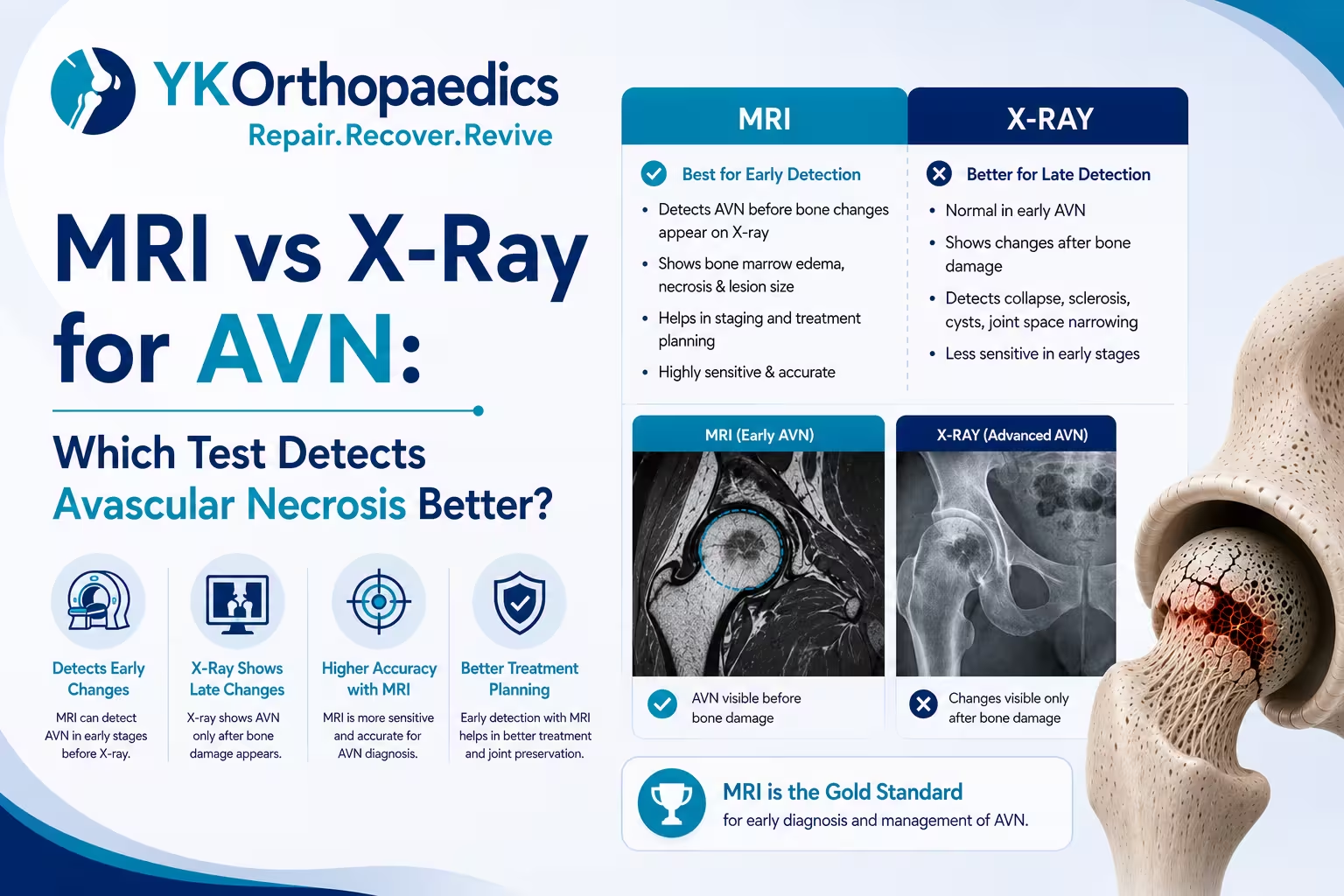
MRI is better for detecting early AVN, while X-ray is useful for identifying established structural damage.
| Feature | MRI | X-Ray |
|---|---|---|
| Detects early AVN | Excellent | May be normal |
| Shows bone marrow changes | Yes | No |
| Shows lesion size/location | Yes | Limited |
| Detects subchondral fracture | Yes | May show later changes |
| Detects femoral-head collapse | Yes | Yes |
| Shows advanced arthritis | Yes | Yes |
| Radiation exposure | No | Yes |
| Cost and availability | Usually higher/less accessible | Usually lower/widely available |
So, if the question is "Which test detects AVN earlier?" — MRI is generally the better test.
Why Is MRI Better for Early AVN?
AVN begins with damage to the bone caused by inadequate blood supply.
During the early phase, the bone may already be affected even though its overall shape remains normal. An X-ray mainly shows the structure and density of the bone, so early AVN may not produce visible changes.
MRI can detect changes within the bone marrow and therefore identify osteonecrosis much earlier.
This makes MRI particularly useful when:
- Hip pain persists but the X-ray is normal
- AVN is suspected because of risk factors
- AVN is already present in the opposite hip
- The doctor needs to assess early disease
- Joint-preserving treatment is being considered
What Can an X-Ray Show in AVN?
X-rays are useful for identifying later structural changes.
Depending on the stage, an X-ray may show:
- Increased bone density
- Areas of bone damage
- Cystic changes
- Subchondral fracture
- Crescent sign
- Femoral-head flattening
- Femoral-head collapse
- Joint-space narrowing
- Secondary osteoarthritis
However, an X-ray can be normal during early AVN.
This is one of the biggest limitations of X-ray when the disease is still developing.
What Can an MRI Show in AVN?
MRI can provide detailed information about the internal condition of the affected bone.
It can show:
- Early bone marrow abnormalities
- AVN lesions
- Serpiginous or geographic lesion borders
- Double-line sign
- Lesion size
- Lesion location
- Weight-bearing involvement
- Subchondral fracture
- Femoral-head collapse
- Cartilage and joint changes
- AVN in the opposite hip
This makes MRI particularly useful for early diagnosis and treatment planning.
MRI vs X-Ray for Different AVN Stages
Stage 1 AVN
MRI: MRI may show AVN-related bone changes even when the X-ray is normal.
X-Ray: The X-ray can appear normal.
Better for early Stage 1 disease: MRI
Stage 2 AVN
MRI: Can show the lesion, its extent and whether collapse has occurred.
X-Ray: May begin showing structural changes but provides less information about the internal extent of the lesion.
Advantage: MRI
Stage 3 AVN
At this stage, structural failure such as a subchondral fracture or femoral-head collapse may have developed.
MRI can show:
- Subchondral fracture
- Crescent sign
- Extent of collapse
- Associated bone changes
X-Ray can show:
- Crescent sign
- Femoral-head collapse
- Flattening
- Structural deformity
Both can be useful at this stage.
Stage 4 AVN
Advanced AVN may involve:
- Femoral-head collapse
- Flattening
- Deformity
- Joint-space narrowing
- Secondary osteoarthritis
At this stage, X-ray can provide valuable information about the overall joint structure, while MRI may provide additional information about the bone and surrounding tissues.
Can an X-Ray Miss AVN?
Yes, particularly during the early stage.
A patient can have AVN-related changes within the femoral head while the X-ray still appears normal.
For example:
Hip pain → X-ray normal → MRI shows early AVN
This does not necessarily mean the X-ray was performed incorrectly. Early AVN may simply not have produced structural changes that are visible on an X-ray.
Can MRI Detect AVN Before an X-Ray?
Yes.
This is one of the major advantages of MRI.
MRI can detect osteonecrosis before many changes become visible on conventional radiographs.
Therefore, if a patient has significant risk factors or persistent symptoms despite a normal X-ray, the doctor may recommend MRI.
Is MRI Always Necessary If the X-Ray Is Normal?
Not necessarily for every patient.
The decision depends on the clinical situation.
MRI may be particularly useful when:
- Symptoms persist
- AVN is strongly suspected
- The patient has significant risk factors
- There is known AVN in the other hip
- Early disease needs to be ruled out
- The doctor needs to assess the lesion before deciding treatment
A normal X-ray should therefore be interpreted in the context of the patient's symptoms and risk factors.
When Is an X-Ray Enough?
X-ray may be sufficient when the goal is to assess established structural disease, especially when advanced AVN is already suspected.
It can help doctors evaluate:
- Femoral-head shape
- Collapse
- Flattening
- Joint-space narrowing
- Arthritis
- Overall bone alignment
It is also commonly used during follow-up to monitor structural progression.
When Is MRI Recommended for AVN?
MRI may be recommended when:
X-Ray Is Normal but Symptoms Continue
Persistent hip or groin pain with a normal X-ray may require further evaluation.
AVN Is Suspected Early
MRI can identify changes before X-ray abnormalities appear.
The Doctor Needs to Measure the Lesion
MRI provides better information about the size and location of the affected bone.
Treatment Planning Is Required
MRI can help determine whether there is:
- No collapse
- Subchondral fracture
- Femoral-head collapse
- Joint degeneration
These findings can influence treatment decisions.
Which Test Is Better for Measuring AVN Size?
MRI is generally more informative for determining the extent and location of an AVN lesion.
This matters because lesion size and location can influence the risk of femoral-head collapse.
A small lesion away from the weight-bearing surface may have a different prognosis from a large lesion involving the weight-bearing portion.
Which Test Is Better for Detecting Femoral-Head Collapse?
Both MRI and X-ray can show femoral-head collapse.
X-ray can clearly demonstrate changes in the overall shape of the femoral head, particularly in established disease.
MRI can show the underlying bone changes and associated findings, including subchondral fracture and the extent of the lesion.
Therefore, the choice depends on what the orthopedic specialist needs to evaluate.
Which Test Is Better for Detecting a Subchondral Fracture?
MRI can detect a subchondral fracture and associated changes within the bone.
The crescent sign can also become visible on radiographs once structural failure has developed.
If an MRI report mentions:
"Subchondral fracture"
or
"Crescent sign"
the finding should be discussed with an orthopedic specialist because it can indicate progression toward collapse.
Can MRI Show AVN in Both Hips?
Yes.
MRI can identify AVN in both femoral heads, including early disease that may not yet be visible on X-ray.
This is particularly useful because bilateral AVN can occur, and one hip may be at a different stage from the other.
For example:
Right hip: Early AVN
Left hip: Advanced AVN with collapse
The treatment approach may therefore be different for each hip.
Does a Normal X-Ray Mean There Is No AVN?
No.
A normal X-ray does not completely rule out early AVN.
If symptoms or risk factors create a strong suspicion, MRI may be required for further evaluation.
This is particularly important in people with risk factors such as:
- Long-term corticosteroid use
- Excessive alcohol consumption
- Previous hip trauma
- Certain blood disorders
- Certain autoimmune conditions
Does a Positive MRI Mean I Need Hip Replacement?
No.
A positive MRI only means that AVN has been identified.
Treatment depends on:
- Stage
- Lesion size
- Lesion location
- Presence of collapse
- Degree of joint damage
- Symptoms
- Age
- Overall health
Early AVN without collapse
Selected patients may be considered for joint-preserving treatment.
Advanced AVN with collapse and arthritis
Total hip replacement may become the more appropriate treatment when pain and functional limitations are significant.
Can MRI Tell the Exact Stage of AVN?
MRI provides the key imaging information needed for staging, but the final stage should be determined by the orthopedic specialist.
Different staging systems, such as Ficat and ARCO, use somewhat different definitions.
For a simple understanding:
Early AVN → MRI abnormality without collapse
Progressing AVN → Subchondral fracture/collapse
Advanced AVN → Collapse + secondary joint degeneration
The complete MRI findings, rather than one phrase in the report, should be considered.
Why Can MRI and X-Ray Give Different Results?
This can happen, particularly when the disease is early.
For example:
X-ray: No obvious abnormality
MRI: Early femoral-head AVN
This is not necessarily a contradiction.
MRI can detect changes within the bone before the structural changes become visible on X-ray.
If there is a difference between the two reports, your orthopedic specialist should interpret the images together with your symptoms and medical history.
Which Test Should You Get First for Suspected AVN?
There is no single answer for every patient.
In many situations, X-ray may be used as an initial imaging test, while MRI is considered when early AVN is suspected or the X-ray does not adequately explain the symptoms.
If AVN is already strongly suspected, MRI can provide much more detailed information about the disease.
The appropriate test should therefore be decided by your doctor.
What Should You Look for in Your MRI or X-Ray Report?
MRI Report
Look for:
- AVN or osteonecrosis
- Lesion size
- Lesion location
- Weight-bearing involvement
- Serpiginous lesion
- Double-line sign
- Subchondral fracture
- Crescent sign
- Femoral-head collapse
- Cartilage damage
X-Ray Report
Look for:
- Increased bone density
- Cystic changes
- Crescent sign
- Flattening
- Collapse
- Joint-space narrowing
- Osteoarthritis
These terms can help you understand the report, but the final diagnosis and stage should be confirmed by your orthopedic specialist.
What Happens After an MRI Diagnosis of AVN?
Treatment depends on the stage and structural condition of the hip.
Early AVN Without Collapse
Possible approaches include:
- Activity modification
- Protected weight-bearing
- Pain management
- Physiotherapy
- Monitoring
- Joint-preserving treatment in selected patients
AVN With Significant Collapse
Once substantial collapse and joint degeneration develop, joint-preserving treatment becomes less predictable.
Total hip replacement may be considered when pain and functional limitations are significant.
The goal is to relieve pain and restore mobility.
When Should You See an Orthopedic Doctor?
An orthopedic consultation is important if you have:
- Persistent hip or groin pain
- A normal X-ray but ongoing symptoms
- MRI-confirmed AVN
- Increasing difficulty walking
- Hip stiffness
- Subchondral fracture
- Femoral-head collapse
- Bilateral AVN
- A history of long-term steroid use
- Previous hip trauma
Early assessment is particularly important when AVN is suspected because treatment aimed at preserving the natural joint is generally more feasible before significant collapse.
Expert Opinion
According to Dr. Yugal Karkhur, a Best Orthopedic Doctor in Gurgaon, MRI and X-ray have different roles in the diagnosis and management of AVN.
MRI is particularly valuable for early detection, identifying the size and location of the lesion and assessing whether structural changes have begun. X-rays remain useful for evaluating established bone collapse, femoral-head deformity and secondary arthritis.
If an X-ray is normal but symptoms or risk factors raise suspicion of AVN, further evaluation with MRI may be appropriate. Once AVN is confirmed, the treatment decision should be based on the stage, lesion characteristics, presence of collapse and condition of the hip joint.
Conclusion
MRI is generally the better test for detecting AVN at an early stage. It can identify changes inside the bone before they become visible on an X-ray and provides detailed information about lesion size, location, weight-bearing involvement and structural damage.
X-rays remain valuable for detecting established changes, including femoral-head collapse, flattening, deformity and secondary arthritis.
So, if you are asking "MRI vs X-ray for AVN—which is better?", the simplest answer is:
MRI → better for early detection and detailed assessment.
X-ray → useful for established structural damage and follow-up.
If you are looking for a Best Orthopedic Doctor in Gurgaon, Dr. Yugal Karkhur can evaluate your X-ray or MRI findings, assess the stage of AVN and discuss the most appropriate treatment options.
FAQs
Is MRI better than X-ray for detecting AVN?
Yes. MRI is more sensitive for detecting early AVN, while X-rays are particularly useful once structural changes such as collapse or arthritis have developed.
Can an X-ray miss early AVN?
Yes. Early AVN may not be visible on an X-ray. MRI can detect changes within the bone before X-ray abnormalities appear.
Can MRI detect Stage 1 AVN?
Yes. MRI can identify early AVN when an X-ray may still appear normal.
What does an X-ray show in advanced AVN?
An X-ray can show femoral-head flattening, collapse, deformity, joint-space narrowing and secondary osteoarthritis.
Can MRI show femoral-head collapse?
Yes. MRI can identify femoral-head collapse and associated findings such as subchondral fracture.
Do I need both MRI and X-ray for AVN?
Not necessarily in every case. Your doctor may use one or both depending on the symptoms, stage and information needed for diagnosis and treatment planning.
Can MRI show the size of AVN?
Yes. MRI can help determine the size and location of the affected bone, which can be important for assessing the risk of progression.
Does a normal X-ray rule out AVN?
No. A normal X-ray does not completely exclude early AVN, particularly when symptoms or risk factors suggest that further investigation is needed.
Does a positive MRI mean I need hip replacement?
No. Treatment depends on the stage, lesion size and location, presence of collapse, joint condition and symptoms.
Which test is best for early AVN?
MRI is generally the preferred imaging test for detecting early AVN because it can identify bone changes before they become apparent on X-ray.