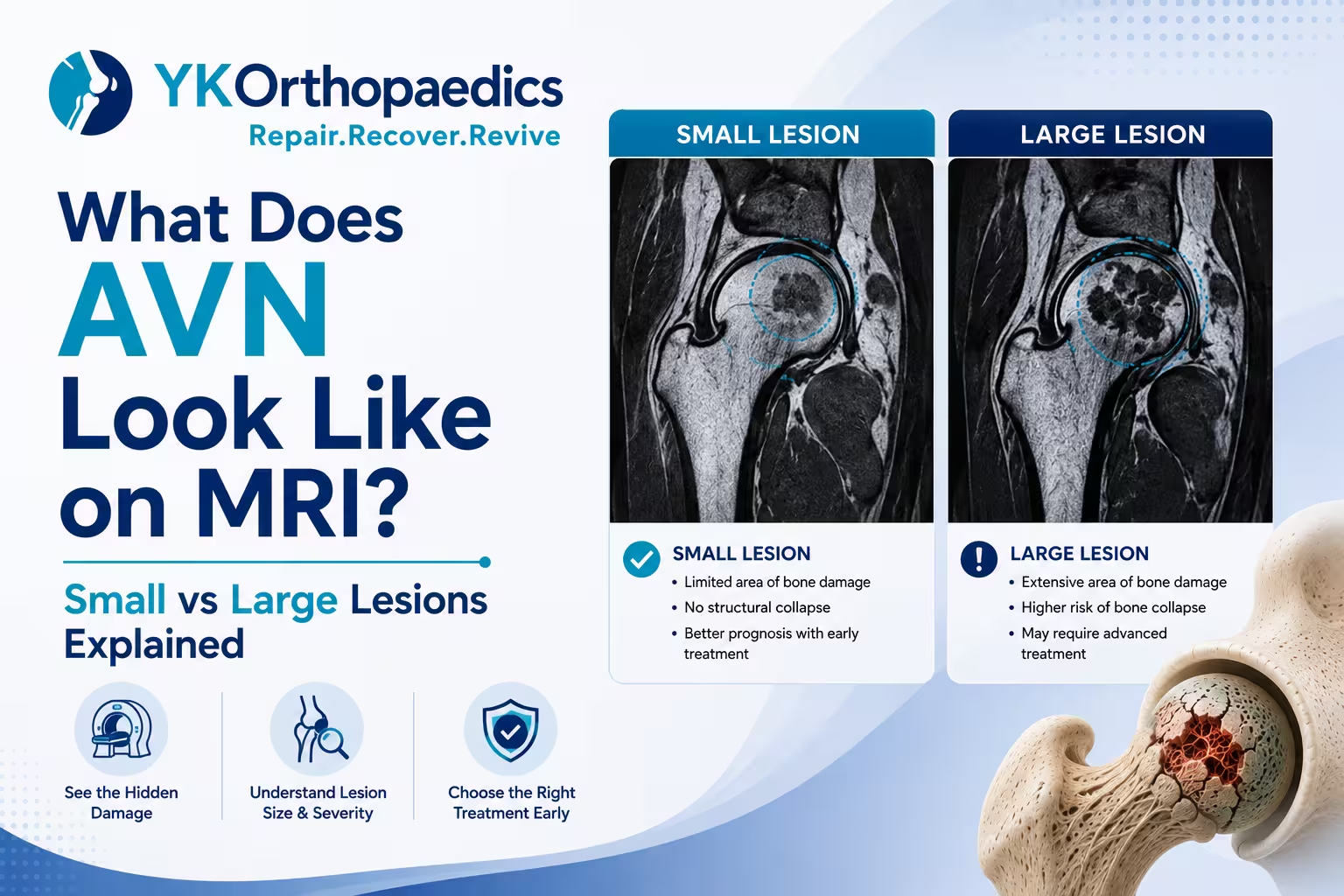
Avascular Necrosis (AVN) has characteristic appearances on MRI, which can help doctors identify the disease at an early stage, determine the size and location of the affected area and assess whether the femoral head has started to collapse. MRI is particularly valuable because it can detect osteonecrosis before it becomes visible on an X-ray and can show the extent of bone damage.
On an MRI, AVN may appear as a geographic or serpiginous area of abnormal bone marrow signal, often with a characteristic rim around the affected bone. The report may also mention terms such as double-line sign, subchondral fracture, crescent sign or femoral-head collapse. The size and location of the lesion are important because small lesions, particularly those outside major weight-bearing areas, may remain stable or heal, while larger lesions involving weight-bearing regions have a greater risk of collapse.
Quick Answer
On MRI, AVN typically appears as an abnormal area within the bone with a characteristic serpiginous/geographic border. The most important things to look for are:
- Small or large lesion
- Location of the lesion
- Whether the weight-bearing area is involved
- Whether there is a subchondral fracture
- Whether the femoral head has collapsed
- Whether there are signs of secondary arthritis
A small AVN lesion without collapse generally has a better prognosis than a large lesion involving the weight-bearing portion of the femoral head.
What Does AVN Look Like on an MRI?
MRI shows changes inside the bone that may not yet be visible on an X-ray.
A typical AVN lesion may be described as:
- Serpiginous
- Geographic
- Well-defined
- Abnormal bone marrow signal
- Rim-like abnormality
- Double-line sign
These MRI findings reflect the boundary between affected bone and the surrounding bone undergoing repair.
MRI is highly sensitive for early osteonecrosis and can sometimes identify AVN before symptoms develop or before X-rays become abnormal.
What Is the "Double-Line Sign" in AVN?
The double-line sign is a classic MRI feature associated with osteonecrosis.
It describes two signal lines forming around the affected area of bone on particular MRI sequences.
If your report says:
"Double-line sign is present."
it supports the diagnosis of AVN.
However, the double-line sign does not by itself tell you whether the AVN is Stage 1, 2, 3 or 4.
The doctor also needs to determine whether there is a subchondral fracture, collapse, arthritis and how much bone is involved.
What Is a Serpiginous Lesion?
A report may say:
"Serpiginous abnormal signal intensity involving the femoral head."
"Serpiginous" simply describes the winding or irregular shape of the boundary of the lesion.
It is a common descriptive term used when reporting osteonecrosis.
The important question is not just that a serpiginous lesion is present, but:
How large is it, where is it located and has the bone collapsed?
What Does a Small AVN Lesion Look Like?
A small AVN lesion involves a relatively limited area of the affected bone.
On MRI, it may appear as a small, well-defined abnormal area with a characteristic rim or serpiginous border.
A small lesion can be particularly favorable when:
- It occupies a limited portion of the femoral head.
- It does not involve the main weight-bearing surface.
- There is no subchondral fracture.
- There is no femoral-head collapse.
- The joint remains structurally preserved.
Small, asymptomatic lesions that are not in major weight-bearing areas may sometimes heal or remain stable.
Does a Small Lesion Mean No Treatment Is Needed?
Not necessarily.
A small lesion may have a better prognosis, but treatment depends on the complete clinical picture.
Your orthopedic specialist may consider:
- Symptoms
- Lesion location
- Risk factors
- Age
- Stage
- Weight-bearing involvement
- Follow-up imaging
Some small lesions can be monitored, while symptomatic or higher-risk lesions may require treatment.
What Does a Large AVN Lesion Look Like?
A large AVN lesion involves a greater portion of the affected bone.
On MRI, it may extend across a substantial portion of the femoral head and may involve the weight-bearing region.
Large lesions are more concerning because the damaged bone has less healthy bone available to support mechanical loads.
According to the Merck Manual, larger infarcts—especially those in weight-bearing areas—have a much greater risk of collapse than small lesions outside major weight-bearing regions.
What Does a Large Lesion Mean for Prognosis?
A large lesion does not automatically mean that collapse has already occurred.
But it can indicate a higher risk of progression.
Therefore, a report saying:
"Large area of AVN involving the weight-bearing portion of the femoral head"
deserves careful orthopedic assessment even if the patient has relatively mild pain.
Small vs Large AVN Lesions
| Feature | Small AVN Lesion | Large AVN Lesion |
|---|---|---|
| Area affected | Limited | Larger portion of bone |
| Weight-bearing involvement | May be absent | More likely |
| Risk of collapse | Generally lower | Generally higher |
| Symptoms | May be mild or absent | May be more significant |
| Prognosis | Often more favorable | More guarded |
| Joint preservation | More feasible in selected cases | May require earlier intervention |
| Monitoring | May be recommended | Often requires closer assessment |
This is a simplified comparison. Lesion location can be as important as lesion size.
Why Does the Location of AVN Matter?
Imagine two people with AVN lesions of similar size.
Person A
The lesion is located away from the main weight-bearing area.
Person B
The lesion involves the weight-bearing portion of the femoral head.
The second lesion may carry a greater risk of structural collapse because it is exposed to repeated mechanical forces during standing and walking.
Therefore, an MRI report should not be interpreted by looking only at the measurement of the lesion.
What Does "Weight-Bearing Area Involved" Mean?
The weight-bearing area is the portion of the femoral head that carries substantial load when you stand and walk.
If AVN affects this region, the weakened bone is exposed to repeated stress.
This can increase the risk that the affected area will eventually deform or collapse.
This is why phrases such as:
"Weight-bearing portion involved"
or
"Superior weight-bearing aspect of the femoral head"
are important in an MRI report.
What Does "No Collapse" Mean on an AVN MRI?
If your MRI says:
"No evidence of femoral-head collapse."
this generally means that significant structural collapse has not been identified.
This is an important finding because treatments aimed at preserving the natural joint are generally more successful before significant collapse develops.
However, "no collapse" does not mean the AVN can simply be ignored.
The size and location of the lesion still matter.
What Does "Subchondral Fracture" Mean?
A subchondral fracture is a fracture just beneath the joint surface.
If your MRI report mentions:
"Subchondral fracture"
or
"Crescent sign"
it indicates that the weakened bone has started to fail structurally.
This is a more advanced finding than AVN without structural collapse.
What Is the Crescent Sign?
The crescent sign represents a subchondral fracture in the affected bone.
It is an important indication that the bone beneath the joint surface has begun to fail.
If the report says:
"Crescent sign noted in the femoral head."
you should discuss the finding with an orthopedic specialist because it can influence both staging and treatment decisions.
What Does Femoral-Head Collapse Look Like on MRI?
When AVN progresses, the weakened portion of the femoral head can collapse.
The MRI may describe:
- Femoral-head collapse
- Flattening
- Irregular contour
- Subchondral fracture
- Loss of spherical shape
- Articular surface deformity
The femoral head normally has a rounded shape. Significant collapse can make the surface irregular and interfere with normal hip mechanics.
Can MRI Show Early AVN Before X-Ray?
Yes.
This is one of the biggest advantages of MRI.
An X-ray can appear normal during the early stages of osteonecrosis, while MRI may already show the affected area.
NIAMS describes MRI as a highly sensitive method for detecting early osteonecrosis, including changes that may appear before X-ray abnormalities.
Therefore:
Normal X-ray + persistent risk factors or symptoms ≠ automatically no AVN.
If clinical suspicion is high, MRI may be recommended.
How Does AVN Look at Different Stages on MRI?
Stage 1 AVN
Typical features may include:
- MRI evidence of early AVN
- Abnormal bone marrow signal
- Characteristic lesion pattern
- No significant collapse
- X-ray may still be normal
This is an early stage where joint-preserving treatment may be considered in selected patients.
Stage 2 AVN
Typical features may include:
- Clearly visible AVN lesion
- Bone structural changes
- No significant femoral-head collapse
- Joint surface relatively preserved
Lesion size and location become particularly important when assessing the risk of progression.
Stage 3 AVN
Typical findings may include:
- Subchondral fracture
- Crescent sign
- Early or established femoral-head collapse
- Change in femoral-head contour
This indicates structural failure of the affected bone.
Stage 4 AVN
Advanced imaging may show:
- Significant femoral-head collapse
- Flattening or deformity
- Cartilage damage
- Joint-space narrowing
- Secondary osteoarthritis
- Other degenerative changes
When extensive collapse and joint degeneration cause significant pain and disability, joint replacement may become the most reliable treatment.
Does Lesion Size Tell You the AVN Stage?
Not by itself.
A large lesion without collapse and a smaller lesion with collapse are very different situations.
For example:
Large AVN + no collapse
→ significant risk may exist, but the joint surface is still preserved.
Small AVN + subchondral fracture
→ structural progression has already occurred.
Therefore, doctors consider lesion size + location + collapse + joint condition, rather than size alone.
How Do Doctors Measure AVN Lesion Size?
Radiologists and orthopedic specialists can estimate the extent of the lesion using MRI and other imaging.
Depending on the staging or measurement method, they may assess:
- Percentage of femoral-head involvement
- Angular extent of the lesion
- Location within the femoral head
- Weight-bearing involvement
The exact measurement method can vary between institutions and staging systems.
You do not need to calculate these measurements yourself from the MRI images.
Can a Large AVN Lesion Be Treated Without Hip Replacement?
Sometimes, if significant collapse has not yet occurred.
Early treatment is important because procedures intended to preserve the natural joint are generally more useful before collapse. Options can include joint-preserving procedures such as core decompression in appropriately selected patients.
However, a large lesion—particularly one involving a weight-bearing area—has a higher risk of progression.
If substantial collapse and degenerative joint changes have already occurred, total hip replacement may become the more reliable option.
Can a Small AVN Lesion Heal on Its Own?
It can happen in selected cases.
Small lesions, especially those outside major weight-bearing areas, may sometimes heal or remain stable.
However, this does not mean every small lesion should simply be ignored.
Your doctor may recommend observation, lifestyle changes, symptom management or other treatment depending on the individual risk profile.
Does a Large AVN Lesion Always Collapse?
No.
A large lesion has a higher risk of collapse, particularly when it involves a weight-bearing area, but MRI findings cannot guarantee that collapse will definitely occur.
The natural history varies between patients.
This is why an orthopedic specialist evaluates the MRI together with symptoms, risk factors and follow-up imaging.
Can AVN Be Present in Both Hips?
Yes.
Non-traumatic AVN can affect both hips, and the two lesions may be different in size or stage.
For example:
Right hip: Small AVN without collapse
Left hip: Large AVN involving the weight-bearing surface
The treatment approach may therefore be different for each hip.
When non-traumatic AVN is identified, both hips are commonly evaluated because disease may be present on the opposite side as well.
What Should You Look for in Your AVN MRI Report?
If you have an MRI report, focus on these phrases:
More favorable findings
- "Small lesion"
- "No subchondral fracture"
- "No femoral-head collapse"
- "Joint surface maintained"
- "No significant degenerative changes"
Findings that require closer assessment
- "Large lesion"
- "Weight-bearing portion involved"
- "Extensive AVN"
- "Subchondral fracture"
- "Crescent sign"
- "Femoral-head collapse"
- "Flattening"
- "Secondary osteoarthritis"
- "Joint-space narrowing"
These phrases provide useful clues, but the complete MRI and clinical assessment are necessary to determine the stage and treatment.
Does MRI Show How Serious My AVN Is?
MRI provides a lot of information, but one finding does not determine the entire severity.
A useful way to think about an AVN MRI is:
1. Is AVN present?
↓
2. How large is it?
↓
3. Where is it located?
↓
4. Is the weight-bearing area involved?
↓
5. Is there a subchondral fracture?
↓
6. Has the femoral head collapsed?
↓
7. Is there arthritis or cartilage damage?
These factors together help determine the prognosis and treatment approach.
What Treatment Is Considered for Small vs Large AVN?
Small, Early Lesion
Depending on the patient, doctors may consider:
- Observation
- Activity modification
- Pain management
- Physiotherapy
- Protected weight-bearing
- Joint-preserving treatment in selected cases
Small lesions outside major weight-bearing areas may sometimes remain stable or heal.
Large or High-Risk Lesion
A larger lesion, particularly one involving the weight-bearing area, may require closer monitoring and earlier consideration of treatment because the risk of collapse is higher.
Collapsed Lesion
If significant collapse and secondary joint degeneration have occurred, total hip replacement may be considered to relieve pain and restore function.
When Should You See an Orthopedic Doctor?
An orthopedic evaluation is particularly important if you have:
- Persistent hip or groin pain
- AVN detected on MRI
- A large AVN lesion
- Weight-bearing surface involvement
- Subchondral fracture
- Crescent sign
- Femoral-head collapse
- Difficulty walking
- Increasing hip stiffness
- A history of steroid use or another AVN risk factor
Early assessment can be important because treatment aimed at preserving the natural joint is generally more effective before significant collapse.
Expert Opinion
According to Dr. Yugal Karkhur, a Orthopedic Doctor in Gurgaon, an AVN MRI should not be judged simply by whether the lesion is described as "small" or "large."
The more important questions are where the lesion is located, whether it involves the weight-bearing portion of the femoral head and whether there is any subchondral fracture or collapse.
A small lesion outside a major weight-bearing area may have a relatively better prognosis, while a large lesion involving the weight-bearing region may carry a higher risk of collapse. Early evaluation can provide more opportunities to consider joint-preserving treatment before significant structural damage occurs.
Conclusion
AVN on MRI typically appears as a characteristic abnormal area within the bone, often with a serpiginous or geographic border. MRI can detect AVN earlier than X-rays and can show the size and location of the lesion as well as whether structural collapse has developed.
A small lesion, especially one outside the major weight-bearing region, may have a better chance of remaining stable or healing. A large lesion involving the weight-bearing portion of the femoral head has a higher risk of progression and collapse.
However, size alone does not determine the AVN stage or treatment. The most important MRI findings include lesion location, weight-bearing involvement, subchondral fracture, femoral-head collapse and secondary arthritis.
If you are looking for a Best Orthopedic Doctor in Gurgaon, Dr. Yugal Karkhur can review your MRI findings, assess the size and location of the AVN lesion and help determine whether joint-preserving treatment or another approach is appropriate.
FAQs
What does AVN look like on MRI?
AVN commonly appears as an abnormal geographic or serpiginous area within the bone, often with a characteristic rim. MRI can detect these changes before they become visible on X-rays.
Is a small AVN lesion dangerous?
A small lesion may have a better prognosis, especially if it does not involve a major weight-bearing area. However, it still requires appropriate assessment and monitoring.
Is a large AVN lesion serious?
A large lesion can be more concerning, particularly when it involves the weight-bearing portion of the femoral head, because it has a greater risk of collapse.
Can a small AVN lesion heal?
Yes, some small lesions—especially those outside major weight-bearing areas—may heal or remain stable.
Does a large AVN lesion always collapse?
No. A large lesion has a higher risk of collapse, but progression varies between patients.
What does "no collapse" mean on an AVN MRI?
It means that significant structural collapse of the femoral head has not been identified. This is generally an important finding when considering joint-preserving treatment.
What does "weight-bearing area involved" mean?
It means the AVN affects the part of the femoral head that carries significant load during standing and walking. Such lesions have a greater risk of structural failure.
What does a crescent sign mean in AVN?
A crescent sign indicates a subchondral fracture and suggests that structural failure has begun in the affected bone.
Can MRI show early AVN when an X-ray is normal?
Yes. MRI is highly sensitive for early osteonecrosis and can identify disease before X-ray abnormalities appear.
Does the size of AVN determine whether I need hip replacement?
No. Treatment depends on lesion size, location, stage, presence of collapse, joint condition, symptoms and other patient-specific factors. Hip replacement is generally considered when significant collapse and joint degeneration have developed.